Radiological Examinations
Accurate imaging is a fundamental cornerstone of modern dental and oral diagnosis. At our clinic, we provide specialized radiological examinations, utilizing state-of-the-art technologies and strictly adhering to quality and safety protocols.
Each examination is selected based on the specific needs of the case, providing the clinical physician with clear and reliable data for accurate diagnosis and treatment planning.
High
radiation protection
We implement international protocols to ensure the safety of our patients.
Unmatched
clarity
We offer radiographs of high diagnostic value.
Radiological
diagnosis
Personalized diagnosis by Maxillofacial Radiologists.
Examination
delivery
Examinations are sent automatically in your preferred format.
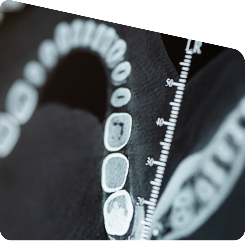
Cone Beam Computed Tomography (CBCT / Dental Scan / CT)
Cone Beam Computed Tomography (CBCT) represents a breakthrough in three-dimensional imaging of the maxillofacial region, offering high accuracy and detail with significantly lower radiation compared to conventional CT scans. It is used for detailed anatomical study, lesion detection, preoperative evaluation, and implant planning.
At our clinic, we perform CBCT scans following the principles of minimal necessary radiation, tailoring the parameters of each examination to the specific needs of the case. The images are rendered with precision and accompanied by a detailed radiological report to optimally support clinical care.
Panoramic Radiography
Panoramic radiography is a fundamental imaging examination that provides a comprehensive view of the upper and lower jaws, teeth, temporomandibular joints, and paranasal sinuses. It serves as a key tool for initial assessment, diagnosis, and planning of dental and maxillofacial surgical treatments.
At our clinic, we use high-resolution digital equipment for panoramic imaging with reduced radiation dose. Imaging is performed with careful attention to proper positioning and technique, ensuring clear images and reliable results for the treating physician.
Lateral Cephalometric Radiography
Lateral cephalometric radiography is an essential diagnostic tool in orthodontic assessment and treatment planning. It provides detailed imaging of the facial skeleton, the relationship between the jaws, and the soft tissues, allowing evaluation of the growth and morphological characteristics of the craniofacial system.
At our clinic, we perform high-resolution digital cephalometric imaging, with the capability for analysis using specialized software. The standardized procedure and careful positioning ensure accuracy and reliability, fully supporting the diagnostic and therapeutic processes of orthodontists and maxillofacial surgeons.
Periapical – Full Mouth Radiography
At our clinic, we perform high-resolution digital cephalometric imaging, with the capability for analysis using specialized software. The standardized procedure and careful positioning ensure accuracy and reliability, fully supporting the diagnostic and therapeutic processes of orthodontists and maxillofacial surgeons.
This method is particularly useful in periodontic and endodontic cases, as well as in preoperative assessment. At our clinic, we apply digital techniques that reduce radiation dose while ensuring excellent imaging clarity, always tailoring the examination to the specific needs of each case.
Posteroanterior Radiography
Posteroanterior (PA) radiography provides a frontal view of the craniofacial skeleton and is primarily used to assess jaw symmetry, monitor orthodontic treatments, and identify pathological findings in the facial skull region. It serves as a complementary examination to cephalometric analysis and is recommended in cases where additional diagnostic information from a frontal projection is required.
At our clinic, posteroanterior imaging is performed using high-precision digital equipment, aiming to minimize radiation exposure while producing diagnostically reliable images. Proper positioning and consistent technique ensure the repeatability and accuracy of the examination.
Bitewing Radiographs
Bitewing radiographs are targeted images used to detect carious lesions on the adjacent surfaces of posterior teeth, as well as to assess bone height in periodontal cases. They provide a clear view of the upper and lower molars and premolars in a single image, allowing early diagnosis even of small lesions.
The examination is performed using digital technology, providing high-resolution images with an exceptionally low radiation dose. It is suitable both for initial screening and for monitoring the progression of caries or periodontal disease with accuracy and consistency in the diagnostic process.
Bitewing Radiography
Bitewing radiography is used to image broader anatomical areas of the upper or lower jaw, providing information that is not easily visible in other intraoral shots. It serves as a useful tool for detecting impacted teeth, foreign bodies, pathological lesions, as well as for assessing fractures or alterations in the anterior region.
The imaging is performed through a simple procedure with minimal radiation dose, utilizing digital technology for clear, easily interpretable images. At our clinic, we apply personalized positioning techniques for each case to achieve optimal imaging and diagnostic accuracy.